
“DGH A” is widely recognized in health technology as a high-precision eye scanning device used in ophthalmology, optometry, and advanced diagnostic imaging. It represents a model or series of devices designed for ocular measurements, imaging, and patient care. Understanding the role and applications of DGH A devices is crucial for healthcare professionals, technicians, and administrators in modern eye care.
What Is the DGH A Eye Scanning Device?
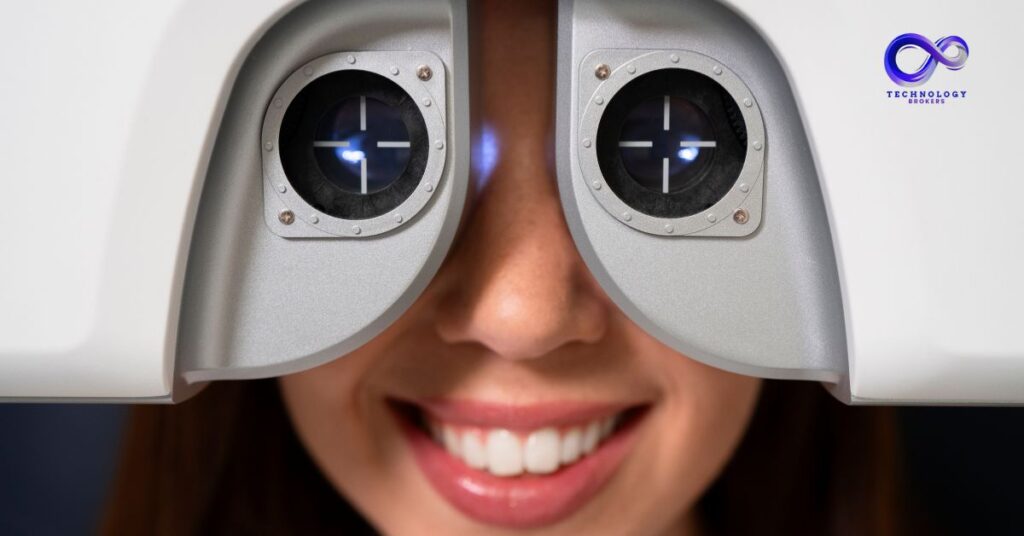
The DGH A device is an ophthalmic diagnostic tool that captures detailed images of the eye, measures ocular structures, and provides critical data for diagnosing eye conditions. It is primarily used for:
- Corneal thickness measurement
- Anterior chamber depth evaluation
- Retinal imaging
- Cataract surgery planning
The device combines precision optics, digital sensors, and advanced software algorithms to deliver highly accurate, reproducible results.
How DGH A Works
The DGH A device utilizes non-invasive scanning technology, often incorporating methods such as:
- Optical coherence tomography (OCT): Generates detailed cross-sectional images of the retina and cornea.
- Ultrasound biometry: Measures axial eye length and anterior chamber depth, essential for intraocular lens calculations.
- Digital imaging: Captures high-resolution images for diagnostic reference and patient records.
The combination of these technologies ensures that clinicians receive accurate anatomical and functional information in real time, improving both diagnostics and treatment outcomes.
Key Features of DGH A Eye Scanning Devices
- High Accuracy: Provides precise measurements of corneal thickness, lens dimensions, and retinal layers.
- User-Friendly Interface: Touchscreen controls, software guidance, and automated calibration make it easy for technicians.
- Non-Invasive and Safe: The scanning process is painless, quick, and safe for all age groups.
- Integration with Health Systems: Can connect with hospital or clinic electronic health record (EHR) systems for seamless data management.
- Compact Design: Suitable for use in clinics, hospitals, and surgical centers.
Applications in Ophthalmology
The DGH A device is critical in eye care diagnostics and surgical planning:
- Cataract Surgery: Measures axial length and anterior chamber depth to determine precise intraocular lens (IOL) power.
- Glaucoma Monitoring: Evaluates corneal thickness and anterior chamber structure to assess intraocular pressure risk.
- Refractive Surgery: Helps plan procedures such as LASIK by mapping corneal curvature and thickness.
- Retinal Health Assessment: Provides high-resolution retinal scans to detect conditions like macular degeneration or diabetic retinopathy.
Advantages of Using DGH A Devices
| Advantage | Description |
| Accuracy | Provides precise anatomical measurements critical for diagnosis and surgery planning |
| Safety | Non-invasive, painless, and suitable for repeated use |
| Efficiency | Quick scans reduce patient wait times and streamline clinic workflows |
| Data Integration | Stores and transmits information to EHR or surgical planning software |
| Versatility | Applicable in routine exams, advanced surgery planning, and research |
Maintenance and Calibration of DGH A Devices
Proper maintenance and regular calibration are essential to ensure the accuracy, reliability, and longevity of the DGH A device. Key practices include:
- Routine Cleaning: Wipe optical lenses, sensors, and surfaces with appropriate cleaning solutions to prevent dust or smudges from affecting scan quality.
- Software Updates: Keep the device software updated to ensure the latest algorithms and features are operational.
- Calibration Checks: Perform calibration checks according to manufacturer guidelines to maintain precise measurements.
- Technician Training: Ensure operators are trained on device handling, scanning techniques, and troubleshooting.
- Service Maintenance: Schedule regular professional maintenance to detect hardware wear or sensor issues early.
Following these steps ensures the device continues to provide high-quality, reliable scans and reduces downtime in clinical settings.
Who Uses DGH A Devices?
- Ophthalmologists: For diagnostic imaging, surgical planning, and patient monitoring.
- Optometrists: For assessing ocular health and refractive measurements.
- Surgical Centers: To ensure accurate pre-operative planning for eye surgeries.
- Research Institutions: For studies involving ocular anatomy, disease progression, or new treatments.
Conclusion
The DGH A eye scanning device is a high-precision, non-invasive diagnostic tool that plays a critical role in modern eye care. By providing accurate measurements and high-resolution imaging, it enhances diagnostic confidence, improves surgical planning, and contributes to better patient outcomes. Its integration into ophthalmology and optometry workflows makes it an essential tool in health technology, ensuring that clinicians can deliver precise, effective, and safe care.
FAQs
Q: Is the DGH A device invasive?
No, it is completely non-invasive and painless for patients.
Q: Can DGH A measure the retina and cornea simultaneously?
Yes, it combines multiple imaging technologies to capture comprehensive ocular data.
Q: Who can operate the DGH A device?
Trained ophthalmic technicians, optometrists, and ophthalmologists can operate it safely.
Q: Is it suitable for children and elderly patients?
Yes, the device is safe for patients of all ages.
Q: Can DGH A integrate with hospital systems?
Yes, it supports electronic health record integration and surgical planning software.